1

Saturday, November 28, 2020

Antibody Titration Anti-A, Anti-B ,Anti-D


PRINCIPLE AND APPLICATIONS:  

Antibody titration is a determination of the concentration of a specific antibody in the patient's serum or to determine the strength of antigen expression on different red cell samples. 

If the concentration of the specific antibody is being determined, the cells must contain the known antigen and the procedure should be performed under the optimal conditions for that antibody. 

The usual applications of titration studies are:

  1. estimating the antibody activity of an alloimmunized pregnant female.
  2. attempting to determine if there is any specificity to an autoantibody
  3. characterizing antibodies that may be high titer, low avidity antibodies

SAMPLE

Either plasma and serum can be titrated.  In the case of a pregnant female, frozen serum or plasma from previous months should be run along with the fresh specimen to determine if there is a rise in titer.

REAGENTS, EQUIPMENT, AND SUPPLIES:

  • Reagent cells known to have the antigen that will be reacting with the titered antibody.  If titering for anti-A or anti-B levels, A1 or B Cells would be used.  If titering for anti-D levels, O cells that are homozygous for D should be used.
  • 12 x 75 mm tubes
  • test tube rack
  • marking pen
  • dispo pipettes
  • physiologic saline
  • serofuge
  • lighted agglutination viewer

PROCEDURE

  1. Label 10 tubes according to the serial dilution: 1, 2, 4, 8, 16, 32, 64, 128, 256, 512 and the patient identification.  The first tube will be undiluted serum.  Tube 2 will be a 1/2 dilution, 4 will be a 1/4 dilution. 
  2. Add 0.3 ml of saline to tubes 2 through 512.  No saline in tube 1
  3. Add 0.3 ml of serum to both tubes 1 and 2.
  4. Use a clean pipette to mix the 1/2 dilution several times and then transfer 0.3 ml to tube 4.
  5. Use a clean pipette to mix the 1/4 dilution several times and then transfer 0.3 ml to tube 8.
  6. Continue the process for all dilutions (512).  Remove 0.3 ml from tube 10 (512) and reserve in a clean tube if the titration needs to be continued.
  7. Label a new set of 10 tubes with the appropriate dilutions.
  8. Using a separate pipettes for each dilution, transfer 2 drops of each tube to the appropriate tubes.
  9. Add one drops of specific red blood cells.
  10. Mix well and test by the appropriate technique for the specific antibody.
  11. Examine test results macroscopically, grade and record the reactions

NOTES AND PRECAUTIONS

  • If titrating anti-A, anti-B, or anti-A,B, the serologic technique is performed by the same method as ABO Typing
  • If titrating Rh, Kell, Duffy, or Kidd antibodies, the serologic technique includes a 37oC followed by antiglobulin testing.
  • If testing a pregnant female, each month serum should be compared to the previous month.
  • Prozone phenomenon may occur so the first tubes may have a weaker reaction than the more diluted serum.  AABB recommends reading the most dilute tubes first and then shake out the other tubes.
  • Careful pipetting is essential.
  • Cells with known antigens may have different reactivity and therefore the serum from each month must use the same cells
  • Measurement is more accurate at larger dilution, therefore the larger dilution should be made before smaller volumes are used to test with the red cells antigen.

INTERPRETATION

  • Observe the highest dilution that produces 1+ macroscopic agglutination
  • The titer is reported as the reciprocal of the dilution level:  32 not 1/32
  • A rise in titer would need to be at least 2 dilution increase between the current specimen and the previous month.
  • For identification of high-titer, low-avidity antibodies would generally have a titer of 64 or greater.

Saturday, November 7, 2020

The National Commission for Allied and Healthcare Professions Bill, 2020 was introduced in Rajya Sabha


                                     Ministry: 
Health and Family Welfare


  • The National Commission for Allied and Healthcare Professions Bill, 2020 was introduced in Rajya Sabha by the Minister of Health and Family Welfare, Dr. Harsh Vardhan, on September 15, 2020.  The Bill seeks to regulate and standardise the education and practice of allied and healthcare professionals.  Key features of the Bill include:


  • Allied health professional: The Bill defines ‘allied health professional’ as an associate, technician, or technologist trained to support the diagnosis and treatment of any illness, disease, injury, or impairment.  Such a professional should have obtained a diploma or degree under this Bill.  The duration of the degree /diploma should be at least 2,000 hours (over a period of two to four years).


  • Healthcare professional: A ‘healthcare professional’ includes a scientist, therapist, or any other professional who studies, advises, researches, supervises, or provides preventive, curative, rehabilitative, therapeutic, or promotional health services.  Such a professional should have obtained a degree under this Bill.  The duration of the degree should be at least 3,600 hours (over a period of three to six years).


  • Allied and healthcare professions: The Bill specifies certain categories of allied and healthcare professions as recognised categories.  These are mentioned in the Schedule to the Bill and include life science professionals, trauma and burn care professionals, surgical and anaesthesia related technology professionals, physiotherapists, and nutrition science professionals.  The central government may amend this Schedule after consultation with the National Commission for Allied and Healthcare Profession.


  • National Commission for Allied and Healthcare Professions: The Bill sets up the National Commission for Allied and Healthcare Professions.  The Commission will consist of: (i) the Chairperson, (ii) Vice-Chairperson, (iii) five members (at the level of Joint Secretary) representing various Departments/ Ministries of the central government, (iv) one representative from the Directorate General of Health Services, (v) three Deputy Directors or Medical Superintendents appointed on a rotational basis from amongst medical institutions including the AIIMS, Delhi and AIIPMR, Mumbai, and (vi) 12 part-time members representing State Councils, among others.


  • Important links:- https://www.aiimsexams.org/
  • https://www.aiims.edu/en.html
  • https://aiimsbhubaneswar.nic.in/
  • https://mohfw.gov.in/

  • Functions of the Commission: The Commission will perform the following functions with regard to Allied and Healthcare professionals: (i) framing policies and standards for regulating education and practice, (ii) creating and maintaining an online Central Register of all registered professionals, (iii) providing basic standards of education, courses, curriculum, staff qualifications, examination, training, maximum fee payable for various categories, and (iv) providing for a uniform entrance and exit examination, among others.
     
  • Professional Councils: The Commission will constitute a Professional Council for every recognised category of allied and healthcare professions.  The Professional Council will consist of a president and four to 24 members, representing each profession in the recognised category.  The Commission may delegate any of its functions to this Council.
     
  • State Councils: Within six months from the passage of the Bill, state governments will constitute State Allied and Healthcare Councils.  The State Councils will consist of: (i) the Chairperson (at least 25 years of experience in the field of allied and healthcare science), (ii) one member representing medical sciences in the state government, (iii) two members representing state medical colleges, (iv) two members representing charitable institutions, and (v) two members from each of the recognised categories of allied and healthcare professions, nominated by the state government, among others.  The State Councils will: (i) enforce professional conduct and code of ethics to be observed by allied healthcare professionals, (ii) maintain respective State Registers, (iii) inspect allied and healthcare institutions, and (iv) ensure uniform entry and exit examinations.
     
  • Establishment of institutions: Prior permission of the State Council will be required to: (i) establish a new institution, or (ii) open new courses, increase the admission capacity, or admit a new batch of students to existing institutions.  If such permission is not sought, then any qualification granted to a student from such an institution will not be recognised under the Bill. 
     
  • Offences and penalties: No person is allowed to practice as a qualified allied and healthcare practitioner other than those enrolled in a State Register or the National Register.  Any person who contravenes this provision will be punished with a fine of Rs 50,000.

Friday, November 6, 2020

Allied and Healthcare Professions Bill 2018

Allied and Healthcare Professions Bill, 2018: A much needed regulation but raises questions on implementation


The allied health professionals in India are not covered by medical or nursing councils. Lab technicians, X-ray technicians, ICU technicians or people who are not covered by any other council and are currently unregulated.

Allied and Healthcare Professions Bill, 2018: A much needed regulation but raises questions on implementation

A big announcement, with perhaps far reaching implications in the Indian healthcare sector, was made on Thursday. The Union Cabinet chaired by Prime Minister Narendra Modi approved the Allied and Healthcare Professions Bill, 2018 for regulation and standardisation of education and services by allied and healthcare professionals.
The bill provides for setting up of an Allied and Healthcare Council of India and corresponding State Allied and Healthcare Councils, which will play the role of a standard-setter and facilitator for professions of Allied and Healthcare. A much needed measure many would say.

The allied health professionals in India are not covered by medical or nursing councils. Think: lab technicians, X-ray technicians, ICU technicians or people who are not covered by any other council and are currently unregulated.
There are over 100 such categories across every branch of healthcare - in eye care for instance: optometrists, in ear problem: audiologists, for diabetes care: diabetologists - covering large number of people across the country.
You would want such critically important professionals to be regulated and adhere to high quality. Since, much of this is still wanting, the plan for new councils for them is arguably much needed. But then this bill has been in the works for over five years now, what is clearly apparent is that this is at best a good starting point.
The bigger challenge lies in effectively implementing it. As every institution teaching these professionals will need to be accredited and those coming under regulation, will fight tooth and nail and given the history of healthcare regulation in India, there could be scope for manipulation.
Also, while the act may lay down strict regulations, what happens when the supply of professionals is less than the demand? How will stipulation on higher qualifications help if supply of talent is not matched? Regulation, after all, works best when the regulators as well as those to be regulated understand the need to be disciplined. Are the regulators always well intentioned?
Ask some private healthcare providers and those within the government in turn point to the various training shops that tend to duck the strict standards and regulations.
After all, this act should not be good only on paper and well drafted piece of regulation but be one that is effectively implemented. And the reason people are sceptical is because there is so much money involved that there is always the danger of abuse. How can that be minimised?
State government and medical community must all be on board and young people joining training courses to be such professionals must see the need for standards - laying down the curriculum, syllabus and laying down the period of training.
How many hospitals today publicly declare their rates and are transparent about their pricing? Also, what if the training provided is not matching and is shoddy?
Every provider, be it a pathology lab that is keen on quality or a diabetics chair that wants to retain its reputation, will need to continue what they do today, which is to largely train their own people.
It all boils down again to history of healthcare regulation in India. It certainly has room for improvement and therefore what is being done to ensure that the new councils will not go down the same path and instead chart out a new course? Also, what will be done to ensure that this will not be used by the big and powerful healthcare providers for eliminating smaller players under the garb of enforcing quality?
What be the view on ensuring high quality delivery in tier II and tier III cities and towns, where even doctors are in short supply, not to talk of high quality allied healthcare professionals? How all these questions get addressed, still needs to be seen.

Friday, November 23, 2018

Ceiling On Tax-Free Gratuity Doubled To Rs. 20 Lakh

The bill moved by Labour Minister Santosh Kumar Gangwar was passed by a voice vote. Lok Sabha had given its approval to the important bill last week.

Ceiling On Tax-Free Gratuity Doubled To Rs 20 Lakh

The Rajya Sabha today passed the Payment of Gratuity (Amendment) Bill without discussion
NEW DELHI:  Parliament today passed a key bill that will empower the government to fix the amount of tax free gratuity and the period of maternity leave with an executive order.

The Rajya Sabha, which has failed to transact any significant businesses in the last fortnight after protests by various parties, today passed the Payment of Gratuity (Amendment) Bill without discussion.

The bill moved by Labour Minister Santosh Kumar Gangwar was passed by a voice vote.

The Lok Sabha had given its approval to the important bill last week.

The legislation will enable the government to enhance the ceiling of tax free gratuity to Rs. 20 lakh from the existing Rs. 10 lakh for employees falling under the Payment of Gratuity Act.

After the implementation of the 7th Pay Commission, the ceiling of gratuity amount for central government employees was doubled to Rs. 20 lakh.
NEW DELHI:  The Gratuity Bill, that empowers the government to fix the amount of tax free gratuity with an executive order, has been passed by Parliament on Thursday.

The bill which was passed by the Lok Sabha last week, was moved by Labour Minister, Santosh Kumar Gangwar in the Rajya Sabha today and passed by a voice vote.

The Payment of Gratuity (Amendment) Bill allows raising the upper limit of payment of tax-free gratuity from Rs. 10 lakh to Rs. 20 lakh. 

Saturday, March 17, 2018

Biochemistry

Biochemistry


Biochemistry, sometimes called biological chemistry, is the study of chemical processes within and relating to living organisms.[1] By controlling information flow through biochemical signaling and the flow of chemical energy through metabolism, biochemical processes give rise to the complexity of life. Over the last decades of the 20th century, biochemistry has become so successful at explaining living processes that now almost all areas of the life sciences from botany to medicine to genetics are engaged in biochemical research.[2]Today, the main focus of pure biochemistry is on understanding how biological molecules give rise to the processes that occur within living cells,[3] which in turn relates greatly to the study and understanding of tissuesorgans, and whole organisms[4]—that is, all of biology.
Biochemistry is closely related to molecular biology, the study of the molecular mechanisms by which genetic information encoded in DNA is able to result in the processes of life.[ Depending on the exact definition of the terms used, molecular biology can be thought of as a branch of biochemistry, or biochemistry as a tool with which to investigate and study molecular biology.
Much of biochemistry deals with the structures, functions and interactions of biological macromolecules, such as proteinsnucleic acidscarbohydrates and lipids, which provide the structure of cells and perform many of the functions associated with life.[6] The chemistry of the cell also depends on the reactions of smaller molecules and ions. These can be inorganic, for example water and metal ions, or organic, for example the amino acids, which are used to synthesize proteins.[7] The mechanisms by which cells harness energy from their environment via chemical reactions are known as metabolism. The findings of biochemistry are applied primarily in medicinenutrition, and agriculture. In medicine, biochemists investigate the causes and cures of diseases.[8] In nutrition, they study how to maintain health wellness and study the effects of nutritional deficiencies.[9] In agriculture, biochemists investigate soil and fertilizers, and try to discover ways to improve crop cultivation, crop storage and pest control.
At its broadest definition, biochemistry can be seen as a study of the components and composition of living things and how they come together to become life, in this sense the history of biochemistry may therefore go back as far as the ancient Greeks.[10] However, biochemistry as a specific scientific discipline has its beginning sometime in the 19th century, or a little earlier, depending on which aspect of biochemistry is being focused on. Some argued that the beginning of biochemistry may have been the discovery of the first enzymediastase (today called amylase), in 1833 by Anselme Payen,[11] while others considered Eduard Buchner's first demonstration of a complex biochemical process alcoholic fermentation in cell-free extracts in 1897 to be the birth of biochemistry.[12][13] Some might also point as its beginning to the influential 1842 work by Justus von LiebigAnimal chemistry, or, Organic chemistry in its applications to physiology and pathology, which presented a chemical theory of metabolism,[10] or even earlier to the 18th century studies on fermentation and respiration by Antoine Lavoisier.[14][15] Many other pioneers in the field who helped to uncover the layers of complexity of biochemistry have been proclaimed founders of modern biochemistry, for example Emil Fischer for his work on the chemistry of proteins,[16] and F. Gowland Hopkins on enzymes and the dynamic nature of biochemistry.[17]
It was once generally believed that life and its materials had some essential property or substance (often referred to as the "vital principle") distinct from any found in non-living matter, and it was thought that only living beings could produce the molecules of life.[25] Then, in 1828, Friedrich Wöhlerpublished a paper on the synthesis of urea, proving that organic compounds can be created artificially.[26] Since then, biochemistry has advanced, especially since the mid-20th century, with the development of new techniques such as chromatography, X-ray diffraction, dual polarisation interferometry, NMR spectroscopy, radioisotopic labeling, electron microscopy, and molecular dynamics simulations. These techniques allowed for the discovery and detailed analysis of many molecules and metabolic pathways of the cell, such as glycolysis and the Krebs cycle (citric acid cycle), and led to an understanding of biochemistry on a molecular level.
Another significant historic event in biochemistry is the discovery of the gene and its role in the transfer of information in the cell. This part of biochemistry is often called molecular biology.[27] In the 1950s, James D. WatsonFrancis CrickRosalind Franklin, and Maurice Wilkins were instrumental in solving DNA structure and suggesting its relationship with genetic transfer of information.[28] In 1958, George Beadle and Edward Tatum received the Nobel Prize for work in fungi showing that one gene produces one enzyme.[29] In 1988, Colin Pitchfork was the first person convicted of murder with DNA evidence, which led to the growth of forensic science.[30] More recently, Andrew Z. Fire and Craig C. Mello received the 2006 Nobel Prize for discovering the role of RNA interference (RNAi), in the silencing of gene expression.

Biomolecules[edit]

The four main classes of molecules in biochemistry (often called biomolecules) are carbohydrateslipidsproteins, and nucleic acids.[33] Many biological molecules are polymers: in this terminology, monomers are relatively small micromolecules that are linked together to create large macromolecules known as polymers. When monomers are linked together to synthesize a biological polymer, they undergo a process called dehydration synthesis. Different macromolecules can assemble in larger complexes, often needed for biological activity.

Carbohydrates[edit]

Glucose, a monosaccharide
A molecule of sucrose (glucose + fructose), a disaccharide
Amylose, a polysaccharide made up of several thousand glucose units
The function of carbohydrates includes energy storage and providing structure. Sugars are carbohydrates, but not all carbohydrates are sugars. There are more carbohydrates on Earth than any other known type of biomolecule; they are used to store energy and genetic information, as well as play important roles in cell to cell interactions and communications.
The simplest type of carbohydrate is a monosaccharide, which among other properties contains carbon, hydrogen, and oxygen, mostly in a ratio of 1:2:1 (generalized formula CnH2nOn, where n is at least 3). Glucose (C6H12O6) is one of the most important carbohydrates; others include fructose (C6H12O6), the sugar commonly associated with the sweet taste of fruits,[34][a] and deoxyribose (C5H10O4). A monosaccharide can switch between acyclic (open-chain) form and a cyclic form. The open-chain form can be turned into a ring of carbon atoms bridged by an oxygen atom created from the carbonyl group of one end and the hydroxyl group of another. The cyclic molecule has an hemiacetal or hemiketal group, depending on whether the linear form was an aldose or a ketose.

In these cyclic forms, the ring usually has 5 or 6 atoms. These forms are called furanoses and pyranoses, respectively — by analogy with furan and pyran, the simplest compounds with the same carbon-oxygen ring (although they lack the double bonds of these two molecules). For example, the aldohexose glucose may form a hemiacetal linkage between the hydroxyl on carbon 1 and the oxygen on carbon 4, yielding a molecule with a 5-membered ring, called glucofuranose. The same reaction can take place between carbons 1 and 5 to form a molecule with a 6-membered ring, called glucopyranose. Cyclic forms with a 7-atom ring called heptoses are rare.
Two monosaccharides can be joined together by a glycosidic or ether bond into a disaccharide through a dehydration reaction during which a molecule of water is released. The reverse reaction in which the glycosidic bond of a disaccharide is broken into two monosaccharides is termed hydrolysis. The best-known disaccharide is sucrose or ordinary sugar, which consists of a glucose molecule and a fructose molecule joined together. Another important disaccharide is lactose found in milk, consisting of a glucose molecule and a galactose molecule. Lactose may be hydrolysed by lactase, and deficiency in this enzyme results in lactose intolerance.
When a few (around three to six) monosaccharides are joined, it is called an oligosaccharide (oligo- meaning "few"). These molecules tend to be used as markers and signals, as well as having some other uses.[36] Many monosaccharides joined together make a polysaccharide. They can be joined together in one long linear chain, or they may be branched. Two of the most common polysaccharides are cellulose and glycogen, both consisting of repeating glucose monomers. Examples are cellulose which is an important structural component of plant's cell walls, and glycogen, used as a form of energy storage in animals.
Sugar can be characterized by having reducing or non-reducing ends. A reducing end of a carbohydrate is a carbon atom that can be in equilibrium with the open-chain aldehyde(aldose) or keto form (ketose). If the joining of monomers takes place at such a carbon atom, the free hydroxy group of the pyranose or furanose form is exchanged with an OH-side-chain of another sugar, yielding a full acetal. This prevents opening of the chain to the aldehyde or keto form and renders the modified residue non-reducing. Lactose contains a reducing end at its glucose moiety, whereas the galactose moiety forms a full acetal with the C4-OH group of glucose. Saccharose does not have a reducing end because of full acetal formation between the aldehyde carbon of glucose (C1) and the keto carbon of fructose (C2).

Lipids[edit]


Structures of some common lipids. At the top are cholesterol and oleic acid.[37] The middle structure is a triglyceride composed of oleoylstearoyl, and palmitoylchains attached to a glycerol backbone. At the bottom is the common phospholipidphosphatidylcholine.[38]
Lipids comprises a diverse range of molecules and to some extent is a catchall for relatively water-insoluble or nonpolarcompounds of biological origin, including waxesfatty acids, fatty-acid derived phospholipidssphingolipidsglycolipids, and terpenoids (e.g., retinoids and steroids). Some lipids are linear aliphatic molecules, while others have ring structures. Some are aromatic, while others are not. Some are flexible, while others are rigid.[39]
Lipids are usually made from one molecule of glycerol combined with other molecules. In triglycerides, the main group of bulk lipids, there is one molecule of glycerol and three fatty acids. Fatty acids are considered the monomer in that case, and may be saturated (no double bonds in the carbon chain) or unsaturated (one or more double bonds in the carbon chain).[40]
Most lipids have some polar character in addition to being largely nonpolar. In general, the bulk of their structure is nonpolar or hydrophobic ("water-fearing"), meaning that it does not interact well with polar solvents like water. Another part of their structure is polar or hydrophilic ("water-loving") and will tend to associate with polar solvents like water. This makes them amphiphilic molecules (having both hydrophobic and hydrophilic portions). In the case of cholesterol, the polar group is a mere -OH (hydroxyl or alcohol). In the case of phospholipids, the polar groups are considerably larger and more polar, as described below.[41]
Lipids are an integral part of our daily diet. Most oils and milk products that we use for cooking and eating like buttercheeseghee etc., are composed of fatsVegetable oils are rich in various polyunsaturated fatty acids (PUFA). Lipid-containing foods undergo digestion within the body and are broken into fatty acids and glycerol, which are the final degradation products of fats and lipids. Lipids, especially phospholipids, are also used in various pharmaceutical products, either as co-solubilisers (e.g., in parenteral infusions) or else as drug carrier components (e.g., in a liposome or transfersome).

Proteins[edit]


The general structure of an α-amino acid, with the amino group on the left and the carboxyl group on the right.
Proteins are very large molecules – macro-biopolymers – made from monomers called amino acids. An amino acid consists of a carbon atom attached to an amino group, —NH2, a carboxylic acid group, —COOH (although these exist as —NH3+ and —COO under physiologic conditions), a simple hydrogen atom, and a side chain commonly denoted as "—R". The side chain "R" is different for each amino acid of which there are 20 standard ones. It is this "R" group that made each amino acid different, and the properties of the side-chains greatly influence the overall three-dimensional conformation of a protein. Some amino acids have functions by themselves or in a modified form; for instance, glutamate functions as an important neurotransmitter. Amino acids can be joined via a peptide bond. In this dehydration synthesis, a water molecule is removed and the peptide bond connects the nitrogen of one amino acid's amino group to the carbon of the other's carboxylic acid group. The resulting molecule is called a dipeptide, and short stretches of amino acids (usually, fewer than thirty) are called peptides or polypeptides. Longer stretches merit the title proteins. As an example, the important blood serum protein albumin contains 585 amino acid residues.[42]

Nucleic acids[edit]


The structure of deoxyribonucleic acid (DNA), the picture shows the monomers being put together.
Nucleic acids, so called because of their prevalence in cellular nuclei, is the generic name of the family of biopolymers. They are complex, high-molecular-weight biochemical macromolecules that can convey genetic information in all living cells and viruses.[2] The monomers are called nucleotides, and each consists of three components: a nitrogenous heterocyclic base (either a purine or a pyrimidine), a pentose sugar, and a phosphate group.[47]

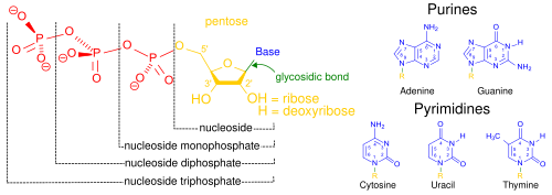
Structural elements of common nucleic acid constituents. Because they contain at least one phosphate group, the compounds marked nucleoside monophosphatenucleoside diphosphate and nucleoside triphosphate are all nucleotides (not simply phosphate-lacking nucleosides).
The most common nucleic acids are deoxyribonucleic acid (DNA) and ribonucleic acid (RNA).[48] The phosphate groupand the sugar of each nucleotide bond with each other to form the backbone of the nucleic acid, while the sequence of nitrogenous bases stores the information. The most common nitrogenous bases are adeninecytosineguaninethymine, and uracil. The nitrogenous bases of each strand of a nucleic acid will form hydrogen bonds with certain other nitrogenous bases in a complementary strand of nucleic acid (similar to a zipper). Adenine binds with thymine and uracil; thymine binds only with adenine; and cytosine and guanine can bind only with one another.
Aside from the genetic material of the cell, nucleic acids often play a role as second messengers, as well as forming the base molecule for adenosine triphosphate (ATP), the primary energy-carrier molecule found in all living organisms.[49] Also, the nitrogenous bases possible in the two nucleic acids are different: adenine, cytosine, and guanine occur in both RNA and DNA, while thymine occurs only in DNA and uracil occurs in RNA.

Metabolism[edit]

Carbohydrates as energy source[edit]

Glucose is an energy source in most life forms. For instance, polysaccharides are broken down into their monomers (glycogen phosphorylase removes glucose residues from glycogen). Disaccharides like lactose or sucrose are cleaved into their two component monosaccharides.

Glycolysis (anaerobic)[edit]

The image above contains clickable links
The metabolic pathway of glycolysis converts glucose to pyruvate by via a series of intermediate metabolites. Each chemical modification (red box) is performed by a different enzyme. Steps 1 and 3 consume ATP (blue) and steps 7 and 10 produce ATP (yellow). Since steps 6-10 occur twice per glucose molecule, this leads to a net production of ATP.
Glucose is mainly metabolized by a very important ten-step pathway called glycolysis, the net result of which is to break down one molecule of glucose into two molecules of pyruvate. This also produces a net two molecules of ATP, the energy currency of cells, along with two reducing equivalents of converting NAD+ (nicotinamide adenine dinucleotide: oxidised form) to NADH (nicotinamide adenine dinucleotide: reduced form). This does not require oxygen; if no oxygen is available (or the cell cannot use oxygen), the NAD is restored by converting the pyruvate to lactate (lactic acid) (e.g., in humans) or to ethanol plus carbon dioxide (e.g., in yeast). Other monosaccharides like galactose and fructose can be converted into intermediates of the glycolytic pathway.[50]

Aerobic[edit]

In aerobic cells with sufficient oxygen, as in most human cells, the pyruvate is further metabolized. It is irreversibly converted to acetyl-CoA, giving off one carbon atom as the waste product carbon dioxide, generating another reducing equivalent as NADH. The two molecules acetyl-CoA (from one molecule of glucose) then enter the citric acid cycle, producing two more molecules of ATP, six more NADH molecules and two reduced (ubi)quinones (via FADH2 as enzyme-bound cofactor), and releasing the remaining carbon atoms as carbon dioxide. The produced NADH and quinol molecules then feed into the enzyme complexes of the respiratory chain, an electron transport system transferring the electrons ultimately to oxygen and conserving the released energy in the form of a proton gradient over a membrane (inner mitochondrial membrane in eukaryotes). Thus, oxygen is reduced to water and the original electron acceptors NAD+ and quinone are regenerated. This is why humans breathe in oxygen and breathe out carbon dioxide. The energy released from transferring the electrons from high-energy states in NADH and quinol is conserved first as proton gradient and converted to ATP via ATP synthase. This generates an additional 28 molecules of ATP (24 from the 8 NADH + 4 from the 2 quinols), totaling to 32 molecules of ATP conserved per degraded glucose (two from glycolysis + two from the citrate cycle).[51] It is clear that using oxygen to completely oxidize glucose provides an organism with far more energy than any oxygen-independent metabolic feature, and this is thought to be the reason why complex life appeared only after Earth's atmosphere accumulated large amounts of oxygen.

Gluconeogenesis[edit]

In vertebrates, vigorously contracting skeletal muscles (during weightlifting or sprinting, for example) do not receive enough oxygen to meet the energy demand, and so they shift to anaerobic metabolism, converting glucose to lactate. The liver regenerates the glucose, using a process called gluconeogenesis. This process is not quite the opposite of glycolysis, and actually requires three times the amount of energy gained from glycolysis (six molecules of ATP are used, compared to the two gained in glycolysis). Analogous to the above reactions, the glucose produced can then undergo glycolysis in tissues that need energy, be stored as glycogen (or starch in plants), or be converted to other monosaccharides or joined into di- or oligosaccharides. The combined pathways of glycolysis during exercise, lactate's crossing via the bloodstream to the liver, subsequent gluconeogenesis and release of glucose into the bloodstream is called the Cori cycle.[52]

Relationship to other "molecular-scale" biological sciences[edit]


Schematic relationship between biochemistry, genetics, and molecular biology.
Researchers in biochemistry use specific techniques native to biochemistry, but increasingly combine these with techniques and ideas developed in the fields of geneticsmolecular biology and biophysics. There has never been a hard-line among these disciplines in terms of content and technique. Today, the terms molecular biology and biochemistry are nearly interchangeable. The following figure is a schematic that depicts one possible view of the relationship between the fields:
  • Biochemistry is the study of the chemical substances and vital processes occurring in living organismsBiochemists focus heavily on the role, function, and structure of biomolecules. The study of the chemistry behind biological processes and the synthesis of biologically active molecules are examples of biochemistry.
  • Genetics is the study of the effect of genetic differences on organisms. Often this can be inferred by the absence of a normal component (e.g., one gene), in the study of "mutants" – organisms with a changed gene that leads to the organism being different with respect to the so-called "wild type" or normal phenotypeGenetic interactions (epistasis) can often confound simple interpretations of such "knock-out" or "knock-in" studies.
  • Molecular biology is the study of molecular underpinnings of the process of replicationtranscription and translation of the genetic material. The central dogma of molecular biology where genetic material is transcribed into RNA and then translated into protein, despite being an oversimplified picture of molecular biology, still provides a good starting point for understanding the field. This picture, however, is undergoing revision in light of emerging novel roles for RNA.[53]
  • Chemical biology seeks to develop new tools based on small molecules that allow minimal perturbation of biological systems while providing detailed information about their function. Further, chemical biology employs biological systems to create non-natural hybrids between biomolecules and synthetic devices (for example emptied viral capsids that can deliver gene therapy or drug molecules).[54]